
Synchrotron radiation methods for studying biological interaction of fibres: an overview
DOI:
https://doi.org/10.13133/2239-1002/18090Abstract
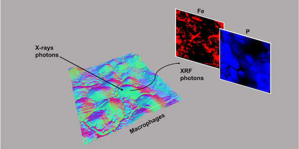
In the last decade, synchrotron-based techniques have emerged as effective tools for investigating biological systems at the sub-cellular level. In asbestos-related studies, X-ray microscopy, combined with X-ray Fluorescence (XRF) and X-ray absorption near edge structure (XANES) enable successfully investigating the interaction between fibres and cells. Microscopy and chemical imaging provide very detailed maps, thus chemically and morphologically characterizing the biological interaction of fibres while monitoring different toxicity mechanisms. Additionally, the combination of microchemical and structural information can clarify metal mobilization processes, highlighting both their intracellular spatial distribution and the possible changes in their valence state induced by the fibres-cells interaction.
The activities within the PRIN 2017-FIBRES project included the utilization of multidisciplinary techniques and expertise to study the interaction of different fibres (chrysotile, erionite and crocidolite) with THP-1-derived macrophages. The numerous experiments carried out at TwinMic (Elettra synchrotron, Trieste, Italy) and ID21 (ESRF, Grenoble, France) beamlines - here discussed and generally presented - offered the opportunity to review the invaluable contribution of synchrotron-based techniques in studying mineral fibres and their biological interaction to step forward in the understanding of asbestos toxicity and carcinogenicity.
Downloads
Published
Issue
Section
License
Copyright (c) 2023 Periodico di Mineralogia

This work is licensed under a Creative Commons Attribution 4.0 International License.
